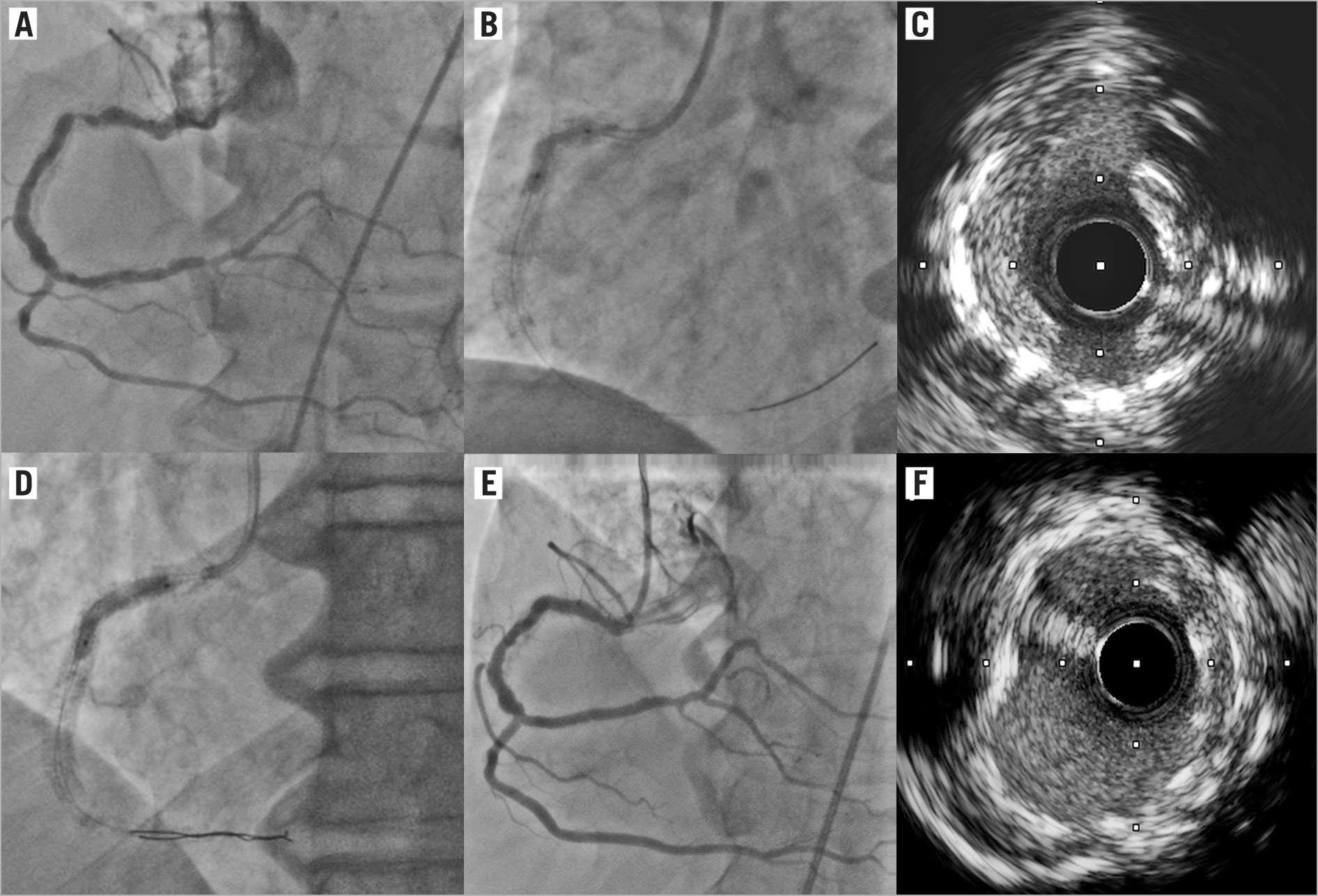
A 59-year-old male with a history of prior (12 years) PCI and drug-eluting stent (DES) implantation on the proximal/mid right coronary artery (RCA) was admitted because of unstable angina. Coronary angiography revealed a focal in-stent restenosis (ISR) at the proximal RCA (Panel A).
Predilatation using a 3.5 mm non-compliant (NC) balloon inflated up to 30 atm was attempted with persistent under-expansion (Panel B). Intravascular ultrasound (IVUS) revealed a heavily calcified neo-intimal plaque at the un-expanded site (Panel C). The lesion was approached by a 3.5×12 mm (sized 1:1 to the reference artery ratio) coronary lithoplasty balloon (LB) (Shockwave IVL, Shockwave Medical, Santa Clara, CA, USA) inflated up to 4 atm before releasing sequential lithotripsy runs at the lesion site, resulting in a full LB and NC (3.5 mm at 24 atm) expansion (Panel D). A new-generation DES (3.5×15 mm) was finally implanted and post-dilated (4.0 mm NC balloon) at the target site obtaining a good final angiographic and IVUS result (Panels E-F).
The use of a coronary LB was recently described as a novel option to facilitate the delivery of interventional equipment and improve stent expansion after modification of calcified plaques in native coronary arteries1. To date very little is known on the performance of the LB for the treatment of undilatable in-stent lesions. Our images highlight the importance of a new technology to overcome the potential limitations of currently available devices (i.e., rotational atherectomy, excimer laser, very high pressure NC balloons) for the treatment of resistant in-stent lesions.
Conflict of interest statement
The authors have no conflicts of interest to declare
