The VBX stent graft (W. L. Gore & Associates) is a useful device. However, there is a possibility of the collapse of a VBX stent that cannot be overcome with endovascular treatment (EVT). Here, we provide the report of a successful attempt at EVT using a self-expanding nitinol stent to recanalise a collapsed and occluded VBX stent in the chronic phase.
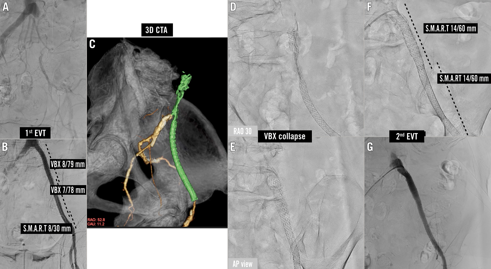
An 84-year-old female was referred to our hospital for bilateral intermittent claudication. The left ankle brachial index (ABI) was 0.49. Computed tomography (CT) and angiography revealed total occlusion of the left iliac artery (Figure 1A). According to intravascular ultrasound (IVUS), the diameter of the proximal target lesion was 8 mm. Two VBX stents (8/78 mm and 7/78 mm) and an S.M.A.R.T stent (8/30 mm) were implanted and post-dilated by an 8 mm non-compliant balloon under intravascular ultrasound guidance (Figure 1B).
Three months later, rest pain recurred and an ischaemic ulcer developed on her left toe, although she was on dual antiplatelet therapy. The ABI was undetectable, and CT and angiography revealed complete collapse of the VBX stent (Figure 1C–Figure 1E, Moving image 1). Although the patient did not have a hunchback posture, she had a habit of bending far forward, which may have greatly influenced the stent deformation. Subsequently, a 14 mm S.M.A.R.T stent (Cordis) was implanted into the VBX stent, just after aspiration and balloon dilatation. This was intended to internally reinforce the VBX stent by taking advantage of the features of the nitinol self-expanding stent (Figure 1F). The final angiogram revealed optimal stent expansion and blood flow. Follow-up CT (Figure 1G) and ultrasound showed excellent patency in the left iliac artery without any stent fracture at one-year follow-up. The oversized self-expanding stents within the VBX stents might contribute to the patency. This case illustrates VBX stent collapse and potential bailout with additional large-diameter self-expanding stent implantation.

Figure 1. Angiographies and CT image. A) Initial angiography of the first EVT. B) Final angiography of the first EVT. C) 3D reconstruction of CT. The collapsed VBX stents are shown in green. D,E) Multidirectional fluoroscopy revealed a collapsed VBX. F) Second EVT. A 14 mm S.M.A.R.T stent was implanted in the collapsed VBX stent. G) Final angiography revealed optimal stent expansion and blood flow. CT: computed tomography; CTA: cardiac computed tomography angiography; EVT: endovascular treatment
Funding
Funding was from institutional sources only.
Conflict of interest statement
The authors report no financial relationships or conflicts of interest regarding the contents of the manuscript.
