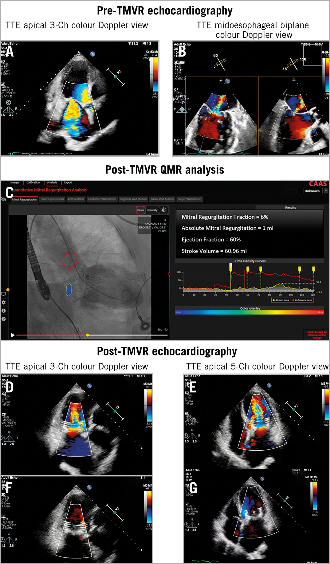
Figure 1. Multi-imaging modality assessment of the mitral valve during TMVR with the HighLife mitral valve. A-B) The transthoracic echocardiography (TTE) 3-Ch colour Doppler view and the transoesophageal echocardiography (TOE) X-plane view show severe mitral regurgitation before TMVR. C) Quantitative angiography of residual mitral regurgitation after TMVR using videodensitometry. D-G) After TMVR, transthoracic echocardiography 3-Ch and 5-Ch views in diastole and systole with colour Doppler shows a well-functioning prosthetic mitral valve. Ch: chamber; QMR: quantitative mitral regurgitation;TMVR: transcatheter mitral valve replacement
Quantitative assessment of aortic regurgitation (AR) using videodensitometric aortography is a well-documented technique1, but the angiographic assessment of mitral regurgitation (MR) is still semiquantitative and operator dependent. The feasibility and accuracy of this novel technology has been investigated and validated in animals2, and the first application in a patient, post-percutaneous mitral replacement, has been reported.
A frail 71-year-old female, hypertensive and diabetic, with persistent atrial fibrillation (AF) and severe MR was admitted with exacerbated shortness of breath. Echocardiography showed severe mitral regurgitation: mitral valve area 3.7 cm², transmitral mean gradient 1 mmHg, mitral valve leaflet length A2/P2 24/11 mm, left ventricular end-diastolic diameter 52 mm, left ventricular end-systolic diameter 40 mm, left atrium 46 mm, left ventricular ejection fraction 47%, and systolic pulmonary pressure 27 mmHg (Figure 1A, Figure 1B, Moving image 1-Moving image 4), with mild-to-moderate aortic regurgitation and mild tricuspid regurgitation. She had multiple previous hospitalisations for heart failure while on optimal medical therapy; transcatheter mitral valve replacement (TMVR) with the novel HighLife (HighLife) transcatheter mitral valve was performed electively after Heart Team consultation34. After successful implantation, a left ventriculogram was obtained in a rotational X-ray (Rx) gantry angulation planned according to a planar projection of a multislice computed topogram that aimed at preventing a 2D fluoroscopic overlap of the left atrium and the ascending aorta, which would preclude the correct videodensitometric assessment5. Using the novel CAAS QMR software (Pie Medical), quantitative angiography analysis of mitral regurgitation (QMR) was conducted at the CORRIB Research Centre for Advanced Imaging and Core Laboratory in Galway, blinded to procedural and echocardiographic data. QMR revealed a regurgitant fraction of 6% and an absolute mitral regurgitation volume of 3 ml, in agreement with the qualitative post-TMVR echocardiographic assessment (Figure 1C-Figure 1G, Moving image 5-Moving image 8).
This case demonstrates the potential of quantitative angiography analysis by videodensitometry in patients undergoing TMVR. A proper fluoroscopic angiographic view derived from multislice computed tomography angiography is essential for an appropriate assessment.
Our first-in-human report included only 1 patient. This first promising experience will be further investigated in more patients undergoing transcatheter mitral valve repair or replacement.
Conflict of interest statement
P.W. Serruys reports consultancy/personal fees from Philips/Volcano, SMT, Novartis, Xeltis, and Meril Life, outside the submitted work. O. Soliman reports grants from NUIG for institutional research. Y. Onuma report grants from NUIG for institutional research, outside the submitted work. The other authors have no conflicts of interest to declare.

