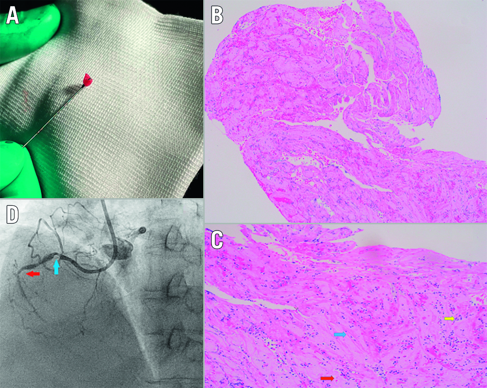
A 65-year-old female with recent non-ST-elevation myocardial infarction underwent high-risk staged percutaneous coronary intervention to her right coronary artery (RCA). Her RCA had significant ostial stenosis followed by a heavy calcified subtotal occlusion of the mid-RCA (Moving image 1). A total of 6,000 units of heparin were given at the start of the procedure. Despite guide extension support, we were unable to pass any balloon across the mid-RCA lesion and hence decided for rotational atherectomy. Three passes with a 1.25 mm ROTABLATOR (Boston Scientific) burr at 180,000 RPM were required to adequately prepare the mid-RCA (Moving image 2). Upon removal of the ROTABLATOR burr, we noticed a piece of red, gelatinous mass wrapped around the burr tip, which was sent for histological evaluation (Figure 1A). We completed the procedure uneventfully by implanting three drug-eluting stents from the ostial to the distal RCA (Moving image 3, Moving image 4).
The histology of this mass showed alternating layers of fibrin, collagen fibres, inflammatory cells and erythrocytes consistent with an organised thrombus (Figure 1B, Figure 1C).
We believe the organised thrombus originated from a hazy segment of the proximal RCA (Figure 1D, blue arrow). Thrombus formation was likely a result of flow stasis from the severe calcific stenosis in the mid-RCA (Figure 1D, red arrow). During atherectomy, the spinning atherectomy burr likely enveloped the proximal RCA thrombus via a centrifugal mechanism, giving it a unique “cotton candy”-like appearance.
These images demonstrate the theoretical concern of using rotational atherectomy in the presence of coronary thrombus, which in turn could lead to complications like distal embolisation or burr entanglement.

Figure 1. Organised coronary thrombus. A) The red gelatinous matter wrapped around the ROTABLATOR burr. B) The cross-sectional histology of the mass showing a tubular, elongated core that shows alternating layers of fibrin and erythrocytes. C) The histology of the mass: a magnified view of the organised thrombus showing the presence of collagen fibres and fibrin (blue arrow), erythrocytes (red arrow) and neutrophils (yellow arrow). D) Coronary angiography: a still image of the RCA angiogram prior to intervention. The blue arrow shows a hazy area in the proximal RCA which is the likely source of the thrombus. The red arrow shows the severe calcific mid-RCA lesion. RCA: right coronary artery
Conflict of interest statement
The authors have no conflicts of interest to declare.
