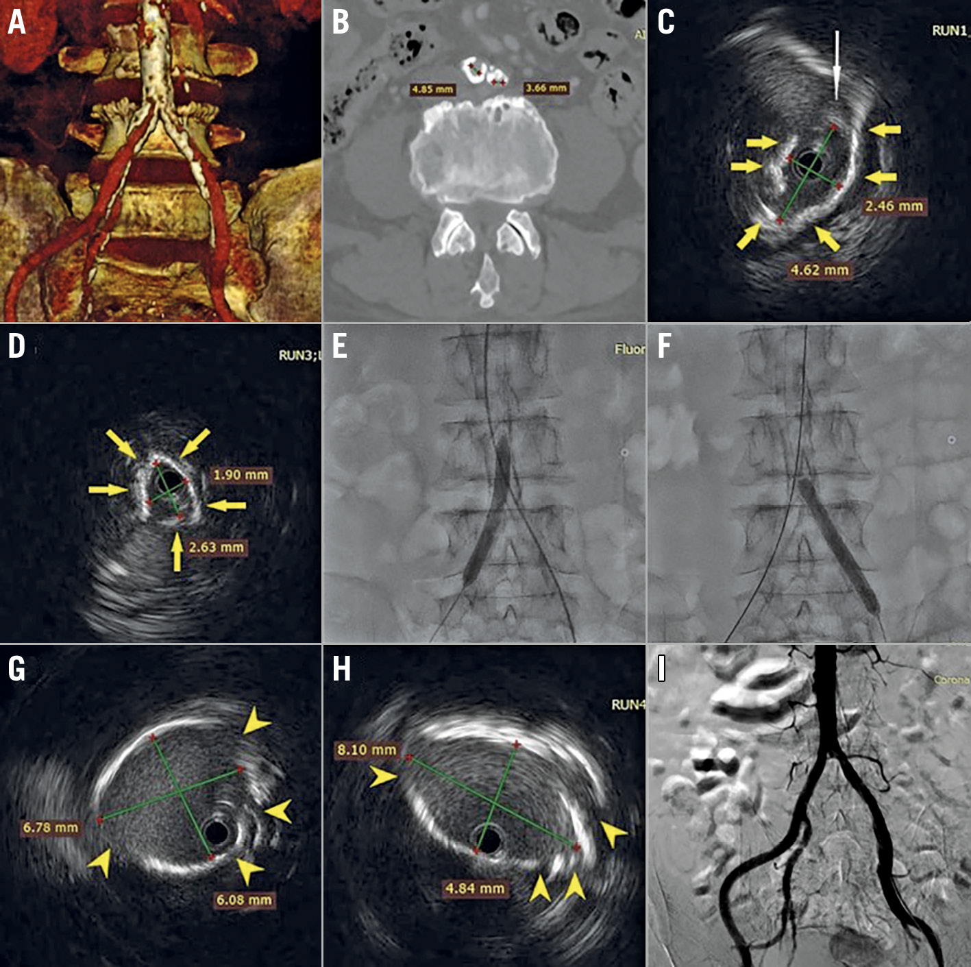
An 80-year-old male with chronic kidney disease (creatinine 200 µmol/L) presented with symptomatic severe aortic stenosis. The computed tomographic scan showed peripheral artery disease (PAD) with a heavily calcified distal aorta, and severely calcified stenoses of the right and left common iliac arteries (CIA), measuring, 4.9 mm and 3.7 mm, respectively (Panel A, Panel B). Transcatheter aortic valve implantation (TAVI) was chosen due to his age and elevated surgical risk. As the patient declined non-femoral approaches, we proceeded with transfemoral TAVI with vessel preparation using Shockwave Intravascular Lithotripsy (IVL) (Shockwave Medical, Santa Clara, CA, USA). IVL enables treatment of calcified stenoses by sonic pressure waves. Both superficial and deep calcium are disrupted with minimal impact on healthy tissue, achieving calcified plaque modification and increasing vessel compliance and size.
The femoral arteries were punctured under ultrasound guidance. A preprocedural aortoiliac angiogram was not performed in order to minimise contrast load and reduce the risk of acute kidney injury. An intravascular ultrasound (IVUS) (OptiCross™ 18 catheter; Boston Scientific, Marlborough, MA, USA), performed to help guide the lithotripsy procedure, confirmed CIA stenoses with napkin-ring calcifications (Panel C – IVUS image of the right common iliac artery, Panel D – IVUS image of the left common iliac artery, yellow arrows denote vessel calcification, white arrow denotes soft plaque). As the non-diseased external iliac artery diameter was approximately 7 mm, a 7×60 mm Shockwave balloon was selected. This was advanced over a 0.014-inch wire to the right CIA and inflated to 4 atm (lithotripsy delivery pressure). Four cycles of 30 pulses each were delivered and then two cycles were delivered to the left CIA (Panel E, Panel F) Repeat IVUS showed calcified plaque fractures with luminal gain (Panel G – IVUS image of the right common iliac artery after IVL, Panel H – IVUS image of the left common iliac artery after IVL, yellow arrowheads denote calcium fractures).
Apart from mild to moderate resistance during insertion of the 14 Fr expandable sheath from the right femoral artery, a 23 mm SAPIEN 3 valve (Edwards Lifesciences, Irvine, CA, USA) crossed the calcified right CIA inside the sheath without much difficulty and was successfully implanted under Sentinel™ (Boston Scientific) cerebral protection. The completion angiography showed no iliofemoral dissection with good flow (Panel I), and the femoral arteriotomy was closed with the ProGlide® (Abbott Laboratories, Abbott Park, IL, USA) pre-closure technique. IVUS-guided IVL can be used to facilitate transfemoral TAVI in patients with calcified PAD and is likely safer than using non-compliant or cutting balloons to dilate severely calcified iliofemoral vessels.
Conflict of interest statement
P. Chiam is a proctor for Edwards Lifesciences and Medtronic. The other authors have no conflicts of interest to declare.
